



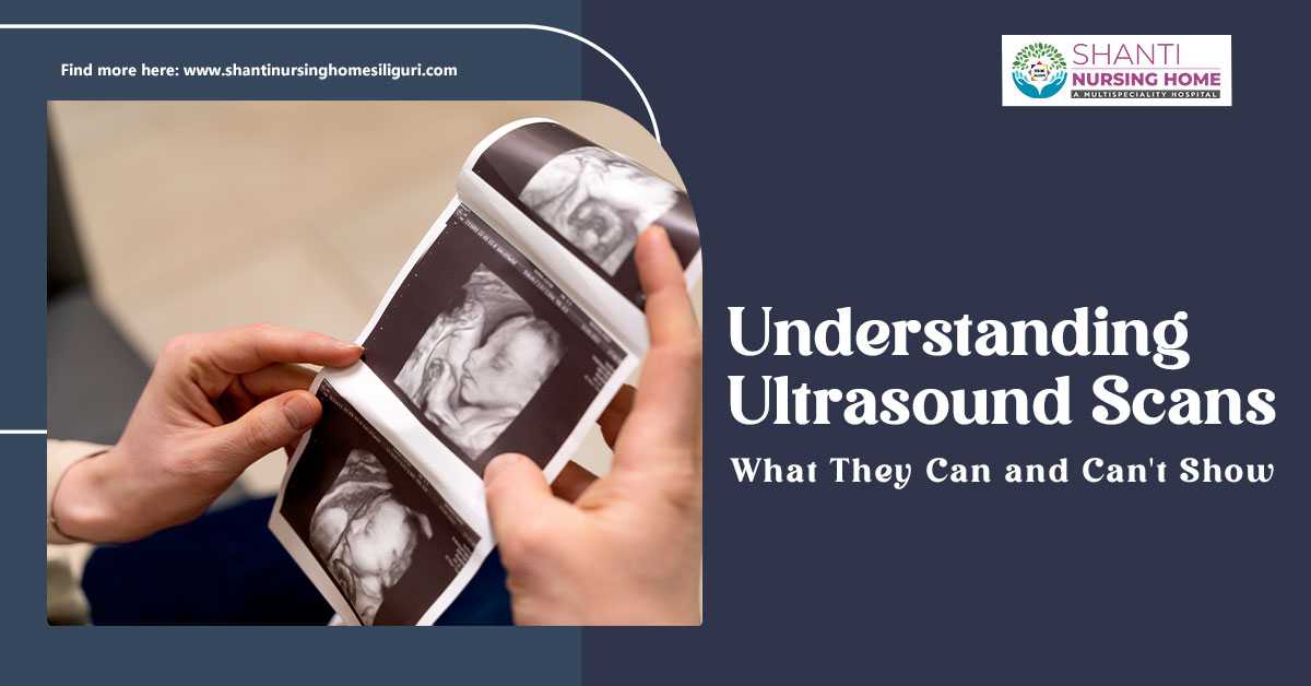

Ultrasound technology uses safe sound waves to create live images of soft tissues, organs, and developing babies.
While they excel at spotting fluid-filled cysts and blood flow, these waves cannot penetrate dense bone or trapped stomach gas.
To overcome these limitations and achieve full-body clarity, doctors frequently pair ultrasounds with CT scans and MRIs.
Ultimately, understanding what this technology can and cannot capture helps set clear medical expectations when choosing the best hospital in Siliguri for your diagnostic needs.
This technology is known as sonography and relies on a handheld device called a transducer to emit sound waves that bounce off internal structures.
Keep reading to understand what ultrasound scans are and what they can and cannot show.
What is an Ultrasound Scan?
An ultrasound scan, also known as sonography, is a safe, non-invasive imaging test that uses high-frequency sound waves to create real-time pictures or videos of the following things.
- Organs
- Tissues
- Blood flow inside your body.
It is ideal for monitoring pregnancy and examining soft tissue because it does not use ionizing radiation.
How Does an Ultrasound Scan Work?
The sole purpose of an ultrasound scan is to create real-time pictures of the inside of the body.
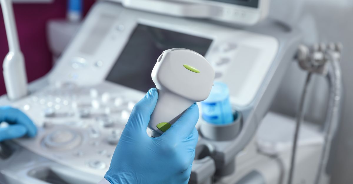
This is done with the help of a handheld probe, called a transducer, which then sends inaudible sound waves into the tissues, which further bounce back as echoes.
After this, a computer then translates these echoes into visuals, allowing doctors to observe the following.
- Organs
- Blood Flow
- Fetal development safely without radiation.
An ultrasound works like a bat hunting in the dark or a submarine navigating underwater.
The following is how an ultrasound scan works.
1. Emission of Sound:
An ultrasound scan works by emitting sound waves, which are completely safe, to create real-time images.
Sound waves are generated by a device called a transducer. Inside, piezoelectric crystals convert applied electrical currents into mechanical vibrations, which compress the surrounding medium to create sound.
These vibrations are used to generate high-frequency sound waves, which are typically between 2 and 18 megahertz and are completely inaudible to human ears.
2. Gel Application:
The whole process of performing an ultrasound starts with the application of a gel on your stomach. To create an airtight seal, a specialized, water-based gel is applied to the skin.
This prevents air pockets, through which sound waves travel poorly, ensuring a clear signal.
3. Receiving Echo:
As the sound waves travel through your body, they hit various tissues, fluids, and bones.
Because different tissues like the muscle, fat, and fluid have different densities, they reflect the sound waves at varying speeds and strengths.
Now, the transducer captures these returning echoes.
4. Processing the Image:
The transducer then converts the returning echoes back into electrical signals. A central processing unit (CPU) calculates how long the echoes took to return and how strong they are, building a map of the internal structures.
This map is instantly displayed as a real-time sonogram on a monitor.
What is an Ultrasound Scan Used For?
Healthcare providers utilize ultrasound exams for three primary purposes, which are as follows.
- Monitoring pregnancy
- Diagnosing medical conditions
- Guiding medical procedures.
These are further described below.
1. For Monitoring Pregnancy: Obstetric Ultrasound
The most common use of an ultrasound scan is for monitoring pregnancy, which is also known as obstetric ultrasound.
In short, this is the most well-known use of ultrasound, and doctors use it for the following purposes.
- Confirm pregnancy and estimate the due date
- Check fetal growth, movement, heart rate, and development
- Screen for abnormalities in the fetus’s developing organs and bones
- Examine the placenta and measure amniotic fluid levels
- Identify multiple features or multiple pregnancies, like twins or triplets.
These are the purposes of performing an ultrasound in the category of monitoring pregnancy.
2. For Diagnosing Medical Conditions
The second reason or purpose of performing an ultrasound scan is to diagnose medical conditions.
These ultrasounds help to look at the internal organs and soft tissues to see if something is wrong, and this happens when a patient experiences symptoms such as.
- Pain
- Swelling
- Infection
It is commonly used to examine the following.
- Evaluate Abdominal Organs:
Ultrasounds are used to evaluate abdominal organs because it evaluates the gallbladder for gallstones, the liver, kidneys for kidney stones, the pancreas, and the spleen.
- Evaluate the Heart, Known as an Echocardiogram:
It is also used to evaluate the heart, which is also known as an echocardiogram. It does so by visualizing the heart valves, chambers, and how effectively the heart is pumping blood.
- Evaluating the Blood Vessels, Known as Doppler Ultrasound:
It measures blood flow and detects blockages, narrowing of arteries, or deep vein blood clots. And this is how ultrasounds are used to evaluate blood vessels, also known as Doppler ultrasounds.
- Check the Pelvic Organs:
Ultrasounds also help in checking the pelvic organs by looking at the uterus and ovaries to find cysts or fibroids, or the prostate gland in men.
- Inspect or Evaluate Other Soft Tissues:
It inspects breast lumps to distinguish between fluid-filled cysts and solid tumors, the thyroid gland, eyes, muscles, and tendons.
3. For Guiding Medical Procedures
Because ultrasounds provide live, real-time images, doctors use them to accurately navigate instruments during complex or delicate procedures.
This includes the following.
- Biopsies:
It is used to perform biopsies by guiding a needle to safely collect tissue samples from tumors or organs like the breast, liver, or thyroid.
- Fluid Drainage:
It also helps in fluid drainage by helping to place needles or catheters to drain painful fluid collections or cysts.
- Injections:
Ultrasounds pinpoint exact locations for nerve blocks or joint injections.
What to Expect From an Ultrasound Scan?
An Ultrasound scan takes approximately 15 to 45 minutes, depending on what part of the body is being scanned.
However, you may need to prepare by fasting or arriving with a full bladder for this.
This procedure is generally painless, though internal ultrasounds like a transvaginal scan may cause slight temporary discomfort.
What are the limitations of an Ultrasound?
An ultrasound cannot do the following.
- Penetrate dense bone
- Travel through air or gas
- Definitely diagnose cancer on its own.
This is because the technology relies on high-frequency sound waves bouncing off internal structures.
It is any barrier that completely blocks or scatters these waves, severely limiting what an ultrasound can scan or see.
The primary limitations of what an ultrasound cannot do or scan are grouped below.
Physical & Structural Barriers of an Ultrasound
1. Cannot Evaluate Dense Bones:
Ultrasounds cannot evaluate dense bones because sound waves bounce completely off hard surfaces.
This means ultrasound cannot see inside adult bones or through the adult skull to image the brain.
2. Catch or Identify Air and Intestinal Gas:
These scans cannot catch or identify air or intestinal gas because the sound waves travel poorly through gas.
This makes ultrasound ineffective for scanning air-filled lungs or organs obscured by the bowel.
3. Identifying Deep Body Structures:
It cannot identify deep body structures because the sound waves used for this weaken as they travel.
Highly deep tissues or examinations on patients with a high body mass index (BMI) can result in fuzzy, inconclusive images.
Diagnostic & Tissue Limitations of an Ultrasound Scan
Ultrasound scans, or ultrasound, also have diagnostic and tissue limitations, which are as follows.
- Definitely confirm cancer:
An ultrasound can find a mass and show if it is solid or fluid-filled. However, it cannot conclusively differentiate between a benign (harmless) and malignant (cancerous) tumor, usually requiring a biopsy.
- Microscopic anomalies:
Very small cellular changes, early-stage tissue damage, or microscopic tumors often will not show up.
- Hollow organ linings:
An ultrasound cannot trace microscopic surface issues like stomach ulcers. Tests like an endoscopy are used instead.
Operational Limitations of an Ultrasound
- Provides 100% operator-independent results:
Ultrasound is entirely dependent on the skill of the person holding the probe. If the technician misses an angle, the condition goes undetected, unlike a CT scan.
- Scanning through wound dressings:
Ultrasounds cannot scan through casts or thick dressings, because the probe must make direct contact with your bare skin using gel to transmit sound waves.
Conclusion
An ultrasound scan uses high-frequency sound waves to create real-time images of the body’s insides.
Because it relies on sound rather than radiation, it is a highly safe, painless, and accessible diagnostic tool used across various medical specialties, including cardiology, gynecology, and obstetrics.
For accurate imaging and expert care, trust the best hospital in Siliguri to guide your health journey.
Schedule your ultrasound scan appointment at Shanti Nursing Home to receive precise results from their expert results from their expert medical imaging team.
